3d Canine Anatomy Software 11 Free Download
3d dog anatomy free download - 3D Dog Anatomy, Dog Anatomy: Canine 3D, Dog Anatomy: Canine 3D, and many more programs. Meet the Canine Anatomy 3D software, a virtual dog (German Shepherd) that. 3D Canine Anatomy Software 1.1 Back. 3D Canine Anatomy for Veterinary Students. 3D Dog Anatomy Software for mobile devices. 3D Bovine Anatomy Software - v1.0. Foot Anatomy Animated Tutorial. Comparative Anatomy: What Makes Us Animals - Crash Course Biology #21. Ultrasounding the Adrenals in Dogs. Labeled dog skeleton pictures.
This video has been updated. Watch the new version here: Try EasyAnatomy for free: Welcome to EasyAnatomy, a living, interactive, 3D anatomy study guide. EasyAnatomy provides a means of engaging students outside the classroom, to improve anatomy retention, clinical application, and day one competencies, without increasing course load or contact time.
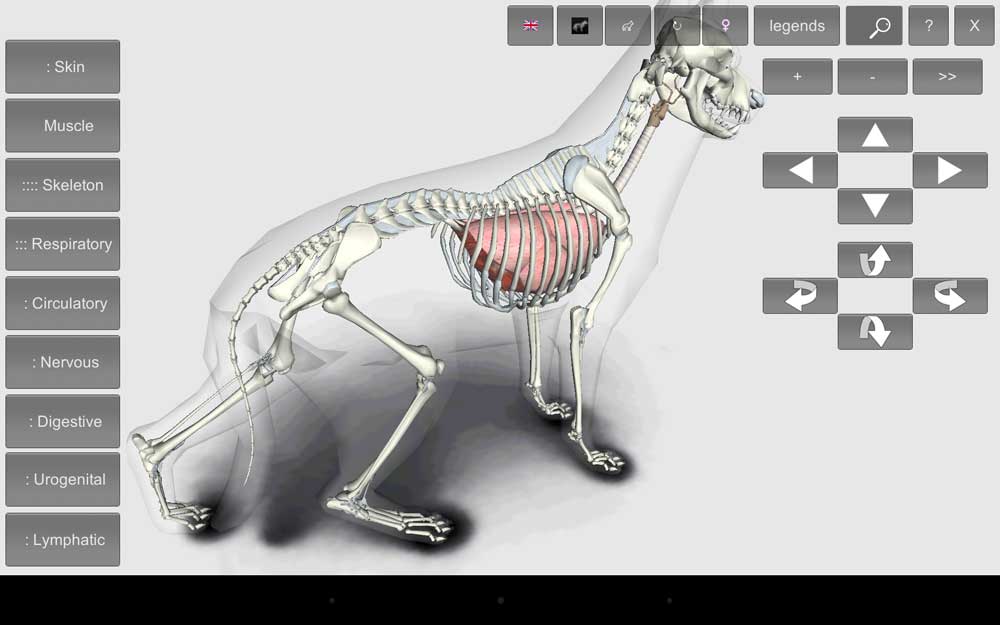
In this video we'll explore EasyAnatomy's interactive 3D canine, the benefits of virtual dissection, and the advantages of adaptive quizzes. EasyAnatomy's immersive learning experience promotes knowledge retention through hands-on discovery and exploration. EasyAnatomy's 21st century dog provides unique anatomical perspective that students can explore from all angles. With EasyAnatomy, the student is in control of the model, they're able to adjust the angle of view, the magnification, and zoom in on the layers which are visible or not visible.
Students can toggle between label tiers and review detailed information written in collaboration with our team of anatomist, to provide clinically relevant and up-to-date content, specific to the individual region's structures and features. EasyAnatomy allows students to see inside without a scalpel, and virtually dissect the canine over and over from any perspective. EasyAnatomy provides views that cannot be achieved with dissection, such as going inside the body cavity while leaving the body wall intact.
Traditionally, most dissection is done with dogs lying on their backs or sides, meaning students are learning some structures in non normal positions. EasyAnatomy's standing model is especially advantageous for the thoracic, abdominal and pelvic regions, and of course you can still move the camera anywhere desired. When studying the muscles of the pelvic limb, students no longer have to cut and reflect superficial muscles to see deeper muscles. With EasyAnatomy you can fade and hide muscles and other structures at will, giving students a more complete view, including muscle origins and insertions. EasyAnatomy's adaptive quizzes reinforce knowledge and provide immediate feedback during self-study, allowing students to retake quizzes and improve their scores over time.
EasyAnatomy's library of a thousand quiz questions written by experienced anatomy professors enables students to choose from a selection of pre-made quizzes or generate their own based on their topic of choice. EasyAnatomy in-app quizzes utilize adaptive learning models, spaced repetition, and confidence-based learning to identify students' weaknesses, to help them work more efficiently during self-directed study. Lan network diagram software. Learn more at EasyAnatomy by LlamaZOO Interactive.

I fully and strongly recommend IVALA for its excellence and accuracy of anatomical details, which enrich the students’ experience inside and outside the dissection rooms in Vet schools around the world. I am a Veterinarian, Veterinary Anatomist and fellow of the World Association of Veterinary Anatomists (WAVA), UK Anatomical Society and of the European Association of Veterinary Anatomists (EAVA). I have been teaching Veterinary Anatomy for 27 years now. In my experience, the most difficult aspect of Anatomy for a student to achieve is to build a 3D visual memory of the organs and how they relate to each other in a 3D space and applying it to a clinical and surgical perspective in different animal species. That is what IVALA brings the best to Vet students and graduates: its innovative approach combines very-detailed high-resolution images obtained from real organs with functional and applied anatomy concepts. With this approach IVALA equips the learner with a plethora of state-of-the-art tools to understand Veterinary Anatomy: a core subject which is pivotal to Veterinary Medicine.